Osteochondritis Dissecans (OCD)
▫️Written by John Keller
✅ Reviewed by Dr. Jenny Hynes on October 25, 2023
Osteochondritis Dissecans (OCD) of the Capitellum is a musculoskeletal anomaly that poses significant threats to joint health and function. Though its name may sound intricate, its implications are profound. Imagine a condition where the bone underneath the cartilage of a joint begins to deteriorate due to insufficient blood flow, leading to potential joint dysfunction and pain (Kijowski, De Smet, & Mukharjee, 2012). Such is the reality for those affected by OCD of the Capitellum.
Keilor Road Physio is a team of physiotherapists who are experts in their field. Book an appointment to see an elbow physio today.
Globally, OCD has been observed to affect a notable portion of the population. In fact, studies have demonstrated that among adolescents, particularly those engaged in regular sporting activities, the incidence rate for OCD can be as high as 29.3 per 100,000 (Kessler et al., 2013). Maruyama et al., (2018), reported the prevalence of OCD lesions between 1% to 3%, notably in adolescent throwing athletes. These numbers stress the importance of being aware of the condition and seeking appropriate intervention when needed.
Neglecting or delaying care for this condition could lead to exacerbated joint damage, pain, and a dramatic decrease in overall joint function. Hence, early detection and intervention are pivotal. Physiotherapy emerges as a cornerstone in this scenario, offering patients not just symptomatic relief but also guiding them towards restoration of function and even potential delay in progression (Takahara et al., 2000).
Decoding Osteochondritis Dissecans (OCD) of the Capitellum
In simple terms, OCD of the Capitellum is a condition where the bone underneath the cartilage in the elbow joint starts to weaken due to inadequate blood flow. As a result, the affected bone and its overlying cartilage can become fragile, sometimes even leading to small fragments breaking off (Edmonds & Polousky, 2013).
If OCD of the Capitellum remains untreated or isn't managed properly, the consequences can be far-reaching. The joint may become stiff and painful, limiting one's ability to move the elbow freely. Over time, this could lead to early-onset arthritis, presenting even more challenges for the individual (Churchill et al., 2016).
The significance of catching OCD of the Capitellum early cannot be emphasized enough. An early diagnosis allows for interventions that can potentially halt or slow down the progression of the disease. More than that, it provides the best chance for preserving joint function and reducing the risk of long-term complications (Matsuura et al., 2014).
Causes and Risk Factors of OCD of the Capitellum
Understanding what leads to a condition is imperative in preventing or managing it. Osteochondritis Dissecans (OCD) of the Capitellum, though complex in nature, has several identifiable causes and risk factors.
Causes:
Trauma: A direct blow or injury to the elbow joint can be a precursor. It can disrupt blood flow to the bone beneath the cartilage, initiating the conditions ripe for OCD to develop (Kusumi et al., 2006).
Repetitive Stress: Like a machine part wearing out with excessive use, the elbow can also succumb to wear and tear. Repetitive actions, especially in sports, can induce incremental damage, eventually leading to OCD of the Capitellum (Stubbs et al., 2001).
Genetic Factors: While it’s still a growing field of research, there are inklings that genetics might play a role, with some families showcasing a history of OCD across generations (Kirsch et al., 2017).
Risk Factors:
Engaging in sports, especially those that involve repetitive throwing or overhead motions, places the elbow under consistent stress. For instance, baseball pitchers, tennis players, and gymnasts often place repetitive loads on their elbow joints, making them more susceptible to this condition (Loughran et al., 2019).
Prevention Insights:
Being proactive when it comes to prevention is the key. Strategies to reduce the risk of OCD include:
Regular Rest: Particularly for athletes, it's vital to intersperse periods of intense activity with adequate rest to allow the elbow joint to recover.
Strength Training: Building the muscles around the elbow can offer additional support and reduce undue stress on the joint.
Proper Technique: In sports, ensuring that one's throwing or movement technique is correct can significantly decrease the risk of repetitive injuries (Matthewson et al., 2015).
Recognising the Signs of OCD of the Capitellum
Osteochondritis Dissecans of the Capitellum manifests in various ways, and early detection can play a pivotal role in effective management.
Characteristic Signs & Symptoms:
Joint Pain: Lateral elbow pain is often the initial indicator. Individuals might experience an aching sensation on the outer part of the elbow, especially post-activity (Glait et al., 2016).
Stiffness: The elbow might feel rigid, often after periods of rest or upon waking.
Limited Range of Motion: Difficulty in bending or straightening the elbow, impeding regular movements.
Clicking Sensations: An unsettling clicking or even a locking sensation during elbow movement can arise (Li & Aspden, 1997).
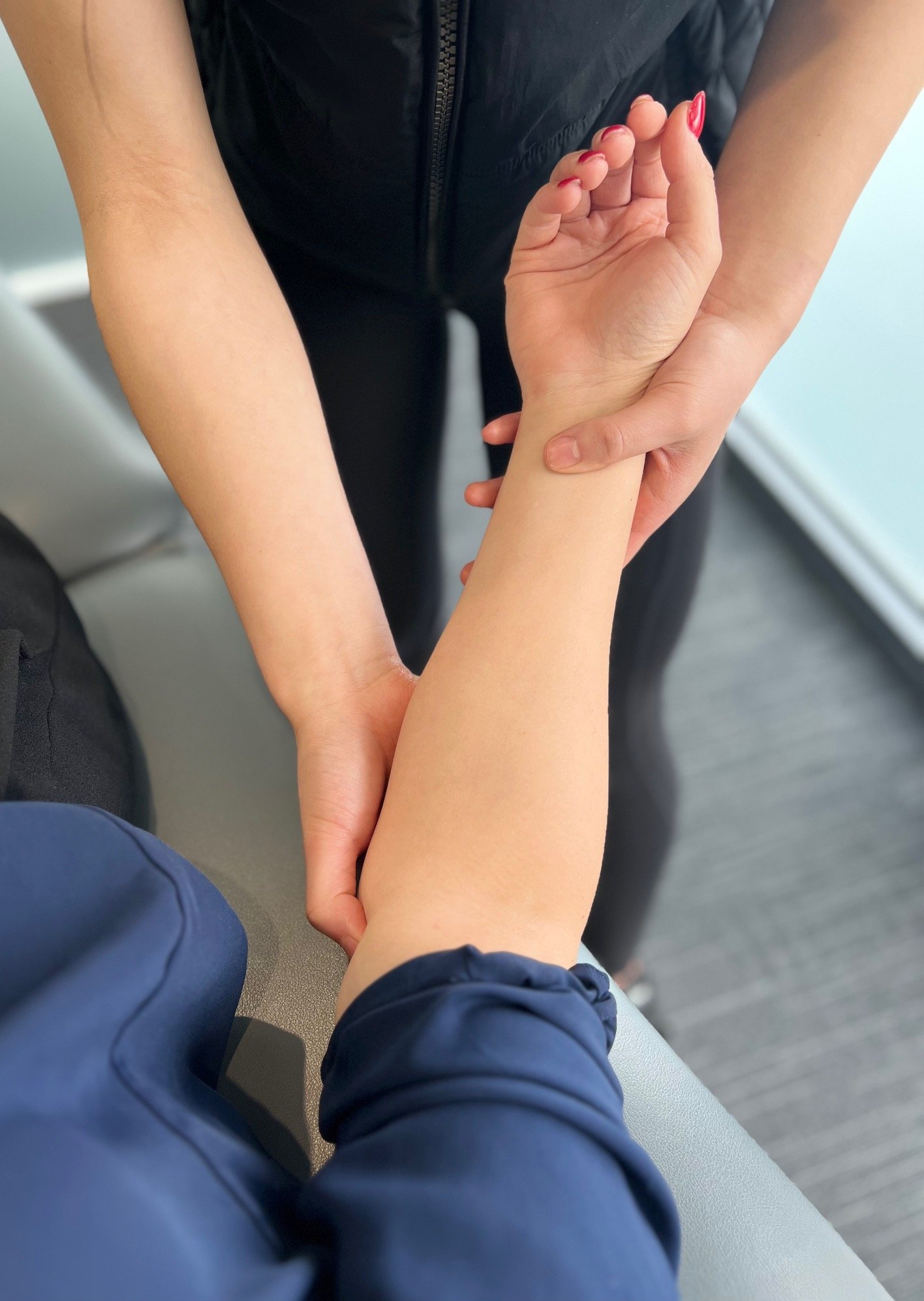
Physical Examination:
At Keilor Road Physiotherapy, we thoroughly assess every elbow injury for potential OCD. This includes looking for any outward signs of change in the elbow including swelling, general movement and special testing.
Two tests we use to help with confirmation include a positive valgus stress test of the elbow. One of our physiotherapists will apply outward (valgus) pressure to the joint while the arm is at a slight angle. Pain or a sense of instability during this test can suggest problems with the inner (medial) part of the elbow, and even deeper issues like OCD (Safran, Ahmad, & Elattrache, 2005).
The milking maneuver is often also positive. The milking maneuver is another elbow ligamentous injury test which evaluates the anterior and posterior bundle of the medial collateral ligament of the elbow. It is performed with the shoulder externally rotated and the elbow flexed beyond 90°. A valgus force is applied by pulling the patient’s thumb while the examiner’s other hand stabilizes the elbow and palpates the medial joint line (Hariri and Safran, 2010). If any of these tests is positive, imaging is indicated and one of our physiotherapists will arrange referral. Imaging could include:
Radiology: X-rays offer a first glance. Specific views, like the anteroposterior (AP) and lateral views, can help identify bone fragments or areas of bone damage. However, early OCD might not be evident on standard X-rays. Research indicates that X-rays have a sensitivity of around 75% in detecting OCD (Richard et al., 2008).
Ultrasound: It provides a real-time imaging method, particularly useful in identifying cartilage irregularities. However, its sensitivity is variable and generally lower than MRI, ranging between 60-70% (Kan et al, 2010).
CT or MRI: These imaging methods provide more detailed visuals. MRI, in particular, can show the exact location and size of cartilage damage, with a sensitivity rate of 90% or higher for OCD detection. CT scans can help visualize bone changes with high detail (Kramer et al., 1993).
Arthroscopy: In certain situations, a clinician may employ arthroscopy, a minimally invasive surgical procedure that allows direct visualization of the joint's interior. This can provide a definitive diagnosis when other methods are inconclusive.
Given the overlap with other elbow conditions, it's paramount not to rely solely on self-diagnosis. Seeking a Keilor Road Physiotherapists expertise ensures accurate identification of the issue and guidance on the optimal management path.

Prevention Strategies for OCD of the Capitellum
Ensuring the elbow joint remains robust requires proactive attention to specific practices and measures.
Strategies for Prevention
Proper Training Techniques: Whether in sports or daily activities, it's imperative to learn and apply correct techniques. For athletes, particularly throwers, mastering the biomechanics of their sport can significantly reduce the risk of undue stress on the elbow joint (Fleisig et al., 1995).
Gradual Progression: Diving into intensive activities without a proper buildup can be a recipe for injury. Instead, individuals should adopt a gradual progression approach, slowly increasing the intensity and duration of their activities to allow the body to adapt (Lyman et al., 2002).
Scheduled Rest Periods: Regular breaks and off-days are essential, especially for those involved in repetitive activities. Periodic rests prevent the buildup of micro-traumas in the joint which, over time, can lead to conditions like OCD (Kerr et al., 2011). Adequate rest and recovery are important factors in preventing overuse injuries. Neglecting this can result in repetitive strain on specific areas, with conditions like OCD of the Capitellum at the elbow being potential outcomes (DiFiori et al., 2014).
Treatment Options for OCD of the Capitellum
Treatment Approaches:
Rest and Activity Modification: Often the initial line of defense, allowing the elbow joint ample time to heal can prove invaluable. Reducing or ceasing aggravating activities provides a natural milieu for recovery (Crawford & Safran, 2006).
Bracing and Immobilization: In some cases, wearing a brace or cast can stabilize the joint, preventing further damage and facilitating healing (Yadao et al., 2004).
Physical Therapy: Engaging in structured physical therapy can enhance joint mobility, reduce pain, and strengthen surrounding muscles. Techniques and exercises are often tailored to the individual's needs and can play a pivotal role in the non-surgical management of OCD.
Surgical Interventions: For severe cases or when non-surgical treatments don't yield the desired results, surgical procedures, like debridement or bone grafting, might be considered. Such interventions aim to restore the integrity of the joint surface and address the damaged bone or cartilage (Takahara et al., 1999).
No two elbows, and by extension, no two cases of OCD, are entirely alike. Therefore, treatment plans should be tailored to the individual, factoring in the severity of the condition, age, activity levels, and personal goals. It's a collaboration between the patient and healthcare professionals to chart the best course forward (Flynn et al., 2004).
As one delves into non-surgical options for managing OCD of the Capitellum, Keilor Road Physiotherapy has a holistic approach that combines evidence-based techniques with personalized care, offering a structured path to manage and potentially overcome the challenges posed by OCD. Our expertise is pivotal in guiding our patients towards optimal joint health and function.
Physiotherapy's Role in Managing OCD of the Capitellum
At the intersection of medical science and rehabilitative therapy, physiotherapy plays a pivotal role in the holistic management of conditions like OCD of the Capitellum. Keilor Road Physiotherapy, with its specialised team and patient-centric approach, offers successful management of OCD,
Keilor Road physiotherapists pride themselves on developing personalised exercise regimens, a cornerstone in the non-surgical management of OCD. Our strategies include:
Addressing Muscle Imbalances: Muscle imbalances, especially around the elbow, can exacerbate joint stress. Through specific exercises, physiotherapists work towards rebalancing these muscles, ensuring optimal force distribution and reduced strain on the joint (Kibler et al., 2012).
Improving Joint Stability: Enhancing the strength and endurance of muscles around the elbow promotes joint stability. Stability exercises aim to protect the affected area from undue stress and prevent further deterioration (Manske & Prohaska, 2008).
Optimising Functional Movement: Whether it's a tennis serve, lifting a kettle, or simply waving hello, functional movement is integral to our lives. Keilor Road physiotherapists focus on restoring and enhancing these movements, ensuring they're efficient and pain-free (Magarey & Jones, 2003).
The journey towards recovery isn't a solitary one; it's a partnership. Keilor Road Physiotherapy places immense value on the collaborative relationship between patients and physiotherapists. Active involvement of the patient in setting goals, understanding their condition, and diligently following through with exercises is paramount. This collaborative approach not only accelerates recovery but also fosters a sense of empowerment among patients (Nicholas et al., 1988).
Restoring Functionality
Physiotherapy is not just about healing; it's about returning individuals to their full potential. For those navigating the challenges of OCD of the Capitellum, physiotherapy can serve as a transformative journey from pain and restriction to freedom and functionality.
Physiotherapy Techniques:
Therapeutic Exercises: These exercises are meticulously designed to strengthen the muscles surrounding the elbow, improve flexibility, and increase joint stability. By progressively enhancing joint function, they play a pivotal role in pain alleviation (Ellenbecker et al., 2009).
Manual Therapy: This hands-on approach involves techniques like joint mobilisations, soft tissue massages, and stretches. Such interventions can reduce joint stiffness, increase range of motion, and enhance blood flow to the affected areas, expediting the healing process (Bialosky et al., 2009).
Neuromuscular Re-education: Our movements, often taken for granted, are orchestrated by intricate neuromuscular coordination. Sometimes, conditions like OCD can disrupt these patterns. Neuromuscular re-education focuses on retraining these movement patterns, ensuring fluidity and efficiency in daily activities (Butler & Moseley, 2003).
Beyond the obvious benefits like pain reduction and improved mobility, physiotherapy aims for holistic wellness. The overarching goal is not just about moving freely but moving with confidence, reducing the risk of recurrence, and truly enhancing the quality of life (Page et al., 2000).
Conclusion
Navigating the Osteochondritis Dissecans (OCD) of the Capitellum may seem daunting, but equipped with knowledge, the journey becomes more than manageable. At its core, OCD of the Capitellum impacts the cartilage and bone of the elbow joint, potentially leading to pain, stiffness, and reduced functionality (Ellenbecker et al., 2009). Factors like trauma, repetitive stress, and genetics can contribute to its onset.
Recognising symptoms early—such as joint pain, limited motion, and characteristic clicking sensations—can pave the way for timely interventions (Magarey & Jones, 2003). These interventions can importantly include proper training techniques, allowing for adequate rest, and adhering to personalised treatment plans, which might range from rest and activity modification to surgical interventions, are paramount (Page et al., 2000).
Keilor Road Physiotherapy can play a key role in seeing you back to 100%. Tailored exercise regimens, manual therapy, and neuromuscular re-education are just a few of the techniques employed to guide patients towards recovery (Butler & Moseley, 2003). Professional guidance cannot be emphasised enough. If you or someone you know grapples with symptoms which might represent OCD of the Capitellum, seeking expert consultation is not just a step towards recovery—it's a leap.
In the face of adversity, remember that with the right support, especially from experienced physiotherapists, a pain-free, functional life isn't a distant dream—it's a tangible reality. Embrace knowledge, seek assistance, and take those proactive strides towards better joint health and a life unhindered.
References
Bialosky, J. E., Bishop, M. D., Price, D. D., Robinson, M. E., & George, S. Z. (2009). The mechanisms of manual therapy in the treatment of musculoskeletal pain: a comprehensive model. Manual therapy, 14(5), 531–538. https://doi.org/10.1016/j.math.2008.09.001
Butler, D. S., & Moseley, G. L. (2003). *Explain pain.* Noigroup Publications.
Churchill, R. W., Munoz, J., & Ahmad, C. S. (2016). Osteochondritis dissecans of the elbow. Current reviews in musculoskeletal medicine, 9(2), 232–239. https://doi.org/10.1007/s12178-016-9342-y
Crawford, D. C., & Safran, M. R. (2006). Osteochondritis dissecans of the knee. The Journal of the American Academy of Orthopaedic Surgeons, 14(2), 90–100. https://doi.org/10.5435/00124635-200602000-00004
DiFiori, J. P., Benjamin, H. J., Brenner, J. S., Gregory, A., Jayanthi, N., Landry, G. L., & Luke, A. (2014). Overuse injuries and burnout in youth sports: a position statement from the American Medical Society for Sports Medicine. *British Journal of Sports Medicine*, 48(4), 287-288.
Edmonds, E. W., & Polousky, J. D. (2013). A review of knowledge in osteochondritis dissecans: 123 years of minimal evolution from König to the ROCK study group. *Clinical Orthopaedics and Related Research*, 471(4), 1118-1126.
Ellenbecker, T. S., Wilk, K. E., Altchek, D. W., & Andrews, J. R. (2009). Current concepts in rehabilitation following ulnar collateral ligament reconstruction. Sports health, 1(4), 301–313. https://doi.org/10.1177/1941738109338553
Fleisig, G. S., Andrews, J. R., Dillman, C. J., & Escamilla, R. F. (1995). Kinetics of baseball pitching with implications about injury mechanisms. The American journal of sports medicine, 23(2), 233–239. https://doi.org/10.1177/036354659502300218
Flynn, J. M., Kocher, M. S., & Ganley, T. J. (2004). Osteochondritis dissecans of the knee. Journal of pediatric orthopedics, 24(4), 434–443. https://doi.org/10.1097/00004694-200407000-00015
Glait, S. A., Rokito, A. S., & Jazrawi, L. M. (2016). Osteochondritis Dissecans of the Capitellum: Diagnosis and Treatment. Bulletin of the Hospital for Joint Disease (2013), 74(1), 37–45.
Hariri S, Safran MR. Ulnar collateral ligament injury in the overhead athlete. Clin Sports Med. 2010;29(4):619–644. [PubMed] [Google Scholar]
Kan, J,H. & Kleinman, P.K. (2010). Pediatric and adolescent musculoskeletal MRI: A case-based approach. Springer Science & Business Media.
Kerr, Z. Y., Collins, C. L., Fields, S. K., & Comstock, R. D. (2011). Epidemiology of player--player contact injuries among US high school athletes, 2005-2009. Clinical pediatrics, 50(7), 594–603. https://doi.org/10.1177/0009922810390513
Kessler, J. I., Nikizad, H., Shea, K. G., Jacobs, J. C., Bebchuk, J. D., & Weiss, J. M. (2013). The demographics and epidemiology of osteochondritis dissecans of the knee in children and adolescents. *The American Journal of Sports Medicine*, 41(2), 320-326.
Kibler, W. B., Sciascia, A., & Wilkes, T. (2012). Scapular dyskinesis and its relation to shoulder injury. The Journal of the American Academy of Orthopaedic Surgeons, 20(6), 364–372. https://doi.org/10.5435/JAAOS-20-06-364
Kijowski, R., De Smet, A. A., & Mukharjee, R. (2012). Magnetic resonance imaging findings in patients with medial epicondylitis. *Skeletal Radiology*, 41(5), 515-519.
Kirsch JM, Thomas JR, Khan M, Townsend WA, Lawton JN, Bedi A.Return to play after osteochondral autograft transplantation of the capitellum: a systematic review. Arthroscopy. 2017;33:1412-20
Kramer J, Recht M, Deely DM, et al. MR appearance of idiopathic synovial osteochondromatosis. J Comput Assist Tomogr. 1993;17:772–776.
Kusumi, T., Ishibashi, Y., Tsuda, E., Kusumi, A., Tanaka, M., Sato, F., Toh, S., & Kijima, H. (2006). Osteochondritis dissecans of the elbow: histopathological assessment of the articular cartilage and subchondral bone with emphasis on their damage and repair. Pathology international, 56(10), 604–612. https://doi.org/10.1111/j.1440-1827.2006.02015.x
Li, B., & Aspden, R. M. (1997). Mechanical and material properties of the subchondral bone plate from the femoral head of patients with osteoarthritis or osteoporosis. Annals of the rheumatic diseases, 56(4), 247–254. https://doi.org/10.1136/ard.56.4.247
Loughran, G. J., Vulpis, C. T., Murphy, J. P., Weiner, D. A., Svoboda, S. J., Hinton, R. Y., & Milzman, D. P. (2019). Incidence of Knee Injuries on Artificial Turf Versus Natural Grass in National Collegiate Athletic Association American Football: 2004-2005 Through 2013-2014 Seasons. The American journal of sports medicine, 47(6), 1294–1301. https://doi.org/10.1177/0363546519833925
Lyman, S., Fleisig, G. S., Andrews, J. R., & Osinski, E. D. (2002). Effect of pitch type, pitch count, and pitching mechanics on risk of elbow and shoulder pain in youth baseball pitchers. The American journal of sports medicine, 30(4), 463–468. https://doi.org/10.1177/03635465020300040201
Magarey, M. E., & Jones, M. A. (2003). Dynamic evaluation and early management of altered motor control around the shoulder complex. *Manual Therapy*, 8(4), 195-206.
Manske, R., & Prohaska, D. (2008). Diagnosis and management of adhesive capsulitis. *Current Reviews in Musculoskeletal Medicine*, 1(3-4), 180-189.
Maruyama, M., Takahara, M., & Satake, H. (2018). Diagnosis and treatment of osteochondritis dissecans of the humeral capitellum. Journal of orthopaedic science : official journal of the Japanese Orthopaedic Association, 23(2), 213–219. https://doi.org/10.1016/j.jos.2017.11.013
Matsuura, T., Suzue, N., Iwame, T., Nishio, S., & Sairyo, K. (2014). Prevalence of Osteochondritis Dissecans of the Capitellum in Young Baseball Players: Results Based on Ultrasonographic Findings. Orthopaedic journal of sports medicine, 2(8), 2325967114545298. https://doi.org/10.1177/2325967114545298
Matthewson, G., Beach, C. J., Nelson, A. A., Woodmass, J. M., Ono, Y., Boorman, R. S., Lo, I. K., & Thornton, G. M. (2015). Partial Thickness Rotator Cuff Tears: Current Concepts. Advances in orthopedics, 2015, 458786. https://doi.org/10.1155/2015/458786
Nicholas, J. A., Rosenthal, P. P., & Gleim, G. W. (1988). A historical perspective of injuries in professional football. Twenty-six years of game-related events. JAMA, 260(7), 939–944.
Page, Phil & Labbe, Andre & Topp, Robert. (2000). Clinical force production of TheraBand® elastic bands. Journal of Orthopaedic and Sports Physical Therapy. 30. A47.
Richard, M. J., Aldridge, J. M., 3rd, Wiesler, E. R., & Ruch, D. S. (2008). Traumatic valgus instability of the elbow: pathoanatomy and results of direct repair. The Journal of bone and joint surgery. American volume, 90(11), 2416–2422. https://doi.org/10.2106/JBJS.G.01448
Safran, M. R., Ahmad, C. S., & Elattrache, N. S. (2005). Ulnar collateral ligament of the elbow. *Arthroscopy: The Journal of Arthroscopic & Related Surgery*, 21(11), 1381-1395.
Stubbs, M. J., Field, L. D., & Savoie, F. H., 3rd (2001). Osteochondritis dissecans of the elbow. Clinics in sports medicine, 20(1), 1–9. https://doi.org/10.1016/s0278-5919(05)70243-x
Takahara, M., Ogino, T., Sasaki, I., Kato, H., Minami, A., & Kaneda, K. (1999). Long term outcome of osteochondritis dissecans of the humeral capitellum. Clinical orthopaedics and related research, (363), 108–115.
Takahara, M., Ogino, T., Takagi, M., Tsuchida, H., Orui, H., & Nambu, T. (2000). Natural progression of osteochondritis dissecans of the humeral capitellum: initial observations. Radiology, 216(1), 207–212. https://doi.org/10.1148/radiology.216.1.r00jl29207
Yadao, M. A., Field, L. D., & Savoie, F. H., 3rd (2004). Osteochondritis dissecans of the elbow. Instructional course lectures, 53, 599–606.

Article by
John Keller
Clinical Director | Sports & Musculoskeletal Physiotherapist
John graduated as a Physiotherapist from the Auckland University of Technology with the John Morris memorial prize for outstanding clinical practise in 2003. John has since completed Post Graduate Diplomas in both Sports Medicine and Musculoskeletal Physiotherapy with distinction, also collecting the Searle Shield for excellence in Musculoskeletal Physiotherapy.

Reviewed by
Dr. Jenny Hynes FACP
Clinical Director | Specialist Musculoskeletal Physiotherapist
Jenny sat extensive examinations to be inducted as a fellow into the Australian College of Physiotherapy in 2009 and gain the title of Specialist Musculoskeletal Physiotherapist, one of only a few physiotherapists in the state to have done so.